F. Ferrari, J. Letsch, L. Morin A. Guignier, L. Marcellin, T. Bourcier
Introduction
It is known that the placement of a pinhole within the cornea results in an increase in depth of field and thus improves near and intermediate visual acuity. This principle has been used since 2011 in the field of corneal refractive surgery through the Kamra® type intracorneal ring technique. This is an additive and reversible surgery whose main advantage is that it does not deteriorate distance vision and allows near vision in emmetropic or ametropic presbyopic patients. Implantation in the pupil area requires mechanical or femtosecond laser-assisted cutting of a LASIK-type cap or the creation of a stromal pocket. The Kamra®inlay is then placed in the bed or pocket around the patient’s previously identified and marked optical axis. This intracorneal implant is made of polyvinylidene fluoride whose biocompatibility has yet to be determined. The external diameter is 3.8 mm and the internal diameter is 1.6 mm. It has numerous micro-perforations designed to facilitate the passage of corneal metabolites (nutrients, electrolytes). The Kamra® implant has undergone successive improvements, including a reduction in thickness (currently five microns) and an increase in the number and distribution of micro-perforations. Although the refractive results seem promising, the limited experience with the use of this biomaterial in the cornea has already led to the observation of certain complications:
- due to the biomaterial: intracorneal iron deposits, diffuse inflammation of the interface, and thinning of the overlying stroma. A confocal microscopy study showed no evidence of intrastromal tissue alteration following placement of a Kamra implant. Another study showed an increase in inlay-induced apoptosis and inflammatory markers in the cornea 24 to 48 hours after surgery with normalization of these parameters six weeks after implantation;
- due to the pinhole effect: loss of visual acuity lines in distance vision, the occurrence of a hypermetropic shift, nocturnal light halos, monocular diplopia;
- due to cutting: dry eyes, loss of visual acuity line.
In addition, the high cost of the implant is a limiting factor for a wider diffusion of this technique whose concept is nevertheless attractive.
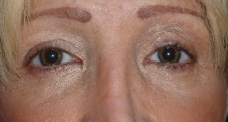
Corneal tattooing is a very old technique, already described by Galen in the 2nd century with the aim of masking a corneal scar or a white cataract by applying walnut or pomegranate bark on a previously cauterized cornea. Later on, Dr. Wecker used Chinese ink by means of non-piercing micro-needles, still in the cosmetic treatment of leukoma. Victor Morax treated the opaque corneas of his patients by introducing a dye into a pre-dissected corneal pocket. More recently, keratopigmentation has been revived for the treatment of iris defects, this time using dyes that meet European standards (Biochromaderm®, Laboratoire BioticPhocea, Marseille, France [SNCH certification no. 0499, sheet 17B, 2011-07-01, Luxembourg]). Thus, in recent years, keratopigmentation has become much “safer” thanks to the use of purified and inert dyes that no longer interact with the surrounding tissues and is increasingly aimed at sighted eyes with iris anomalies.
The combination of the two techniques, intracorneal ring on one hand, and keratopigmentation on the other hand, it seems to us to be at the origin of new corneal surgery techniques, including the treatment of presbyopia. The purpose of our study is to use the keratopigmentation technique to create an intrastromal concentric black (or colored) annular centered on the visual axis, with an internal diameter of slightly less than 2 mm, likely to produce a pinhole effect (Patented technique, U.S. Provisional Application No. 61/713,013). We present here the results of a preliminary feasibility study performed on pig eyes.
Section snippets
Material and methods
Five eyes of pigs were enucleated eight hours before the experiment was used (Strasbourg slaughterhouses). The laser used was a femtosecond laser (Visumax®, Jena, Carl Zeiss®). The eyes were treated with the Intra Corneal Ring (ICR®) program corresponding to the creation of tunnels for intracorneal rings (used for keratoconus and the treatment of moderate myopia). The parameters of the tunnel diameters were modified as follows: internal diameter: 1.8 mm,
Results
Corneal imagery by anterior segment spectral-domain OCT allowed control of the depth and regularity of the stromal cutout serving as a bed for the dye injection, as well as the loss of corneal reflectivity related to the “mask effect” of the pigment. For the control eye, the cutting interface was visualized at a depth of 221 μm (Fig. 4).
A linear hyperreflective blade located at the cutting interface and associated with a posterior shadow cone has been implemented
Discussion
The surgical treatment of presbyopia has progressed considerably in recent years. Several surgical approaches have been successively proposed: central island, decentered island, centered bulging ring, and tilt techniques. In spite of the constant improvement of these techniques, in particular the techniques of tilting with an increase of the depth of field and improvement of the binocular vision, the patients are sometimes embarrassed in distance vision by the visual acuity of
Conclusion
This study represents, to our knowledge, the first experimental attempt to bring together two a priori distant concepts: the creation of an intracorneal pinhole to treat presbyopia on the one hand, and corneal tattooing on the other. The first feasibility study of annular keratopigmentation (PresbyRing®) in post-mortem animals gave encouraging results. It must be confirmed by other studies carried out in animals in vivo and then
Declaration of interest
The authors declare that they have no conflicts of interest in relation to this article.
Scientific article DOI : https://doi.org/10.1016/j.jfo.2013.01.004
References (18)
- J.L. Alió et al. Femtosecond-assisted keratopigmentation for functional and cosmetic restoration in essential iris atrophy J Cataract Refract Surg (2011)
- D. Hirsbein et al. Corneal tattooing for iris defects J Fr Ophtalmol (2008)
- A.K. Dexl et al. Reading performance and patient satisfaction after corneal inlay implantation for presbyopia correction: two-year follow-up J Cataract Refract Surg (2012)
- O. Seyeddain et al. Small-aperture corneal inlay for the correction of presbyopia: 3-year follow-up J Cataract Refract Surg (2012)
- A.K. Dexl et al. Reading performance after implantation of a small-aperture corneal inlay for the surgical correction of presbyopia: two-year follow-up J Cataract Refract Surg (2011)
- A.K. Dexl et al. Central and peripheral corneal iron deposits after implantation of a small-aperture corneal inlay for correction of presbyopia J Refract Surg (2011)
- D.I. Bouzoukis et al. Visual outcomes and safety of a small diameter intrastromal refractive inlay for the corneal compensation of presbyopia J Refract Surg (2012)
- M.R. Santhiago et al. Short-term cell death and inflammation after intracorneal inlay implantation in rabbits J Refract Surg (2012)